A Guide to Understand Teeth with Diagrams
Teeth play a vital role in the human body. It is there for grinding and breaking down food into smaller particles. A tooth anatomy is beneficial for learning about human tooth structure. From this article, here illustrates what does teeth look like, its function, and how to create a tooth diagram online.
1. How Does Teeth Look Like
Teeth play a vital role in the human body. It is there for grinding and breaking down food into smaller particles. The masticated food bolus is pushed towards the esophagus for further digestion. Without the help of teeth, it is not possible to break down food into a digestible form.
It is essential to have sound knowledge about tooth anatomy. Human beings have two sets of teeth on each jaw. The upper jaw and the lower jaw contain an equal number of teeth.
The movable lower jaw is responsible for all kinds of movements of the mouth. It facilitates the chewing of food. With the help of teeth diagrams, it is easier to understand the shape and function of teeth.
Four types of teeth include:
- Incisors: These teeth are further subdivided into central incisors and lateral incisors. These have incisal edges that help to cut food.
- Canines: these teeth have pointed cusps and the longest roots. These are very sharp, and tearing food pieces into smaller bits is their central purpose.
- Premolars: These teeth have 3 to 4 cusps for crushing food particles.
- Molars: These teeth are broad and flat. They contain 4 to 5 cusps and 2 to 3 roots. Molars grind the food into a paste so that it becomes easier to digest and swallow. The third molar tooth in each quadrant of the mouth is known as the wisdom tooth.
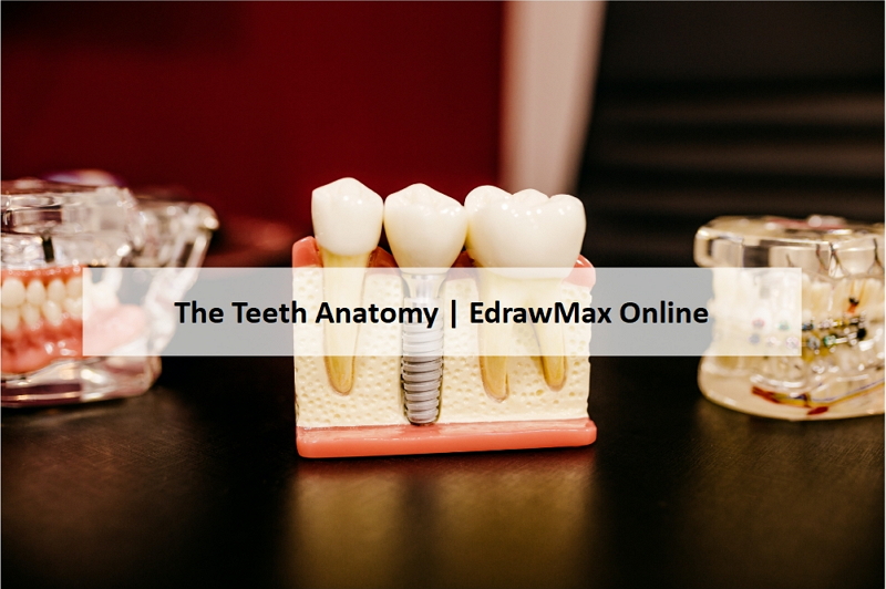
2. The Teeth Anatomy
Teeth are there for masticating food. Improperly masticated food can cause indigestion. The teeth' function depends on the tooth structure. Tooth diagrams are there to teach the teeth anatomy.
The mid-sagittal plane divides the human mouth into four quadrants. Teeth are arranged in two arches in maxillary and mandibular. (Maxillary is the upper jaw, whereas the mandibular is the lower jaw). Children develop deciduous teeth, later replaced by permanent teeth. Human beings have 32 permanent teeth. Even though the permanent dentition contains 32 teeth, many people don’t develop the last molars in each quadrant. In some cases, impacted wisdom teeth cause pain and require dental extraction.
The diagrammatic representation of the jaws helps in understanding the teeth anatomy of human beings. Each quadrant of the jaw consists of:
- 1 Central Incisor
- 1 Lateral incisor
- 1 Canine
- 2 Premolars
- 2-3 Molars
Hence, each quadrant consists of 7- 8 teeth. The incisors are at the front of the mouth. Canines are adjacent to the lateral incisors. The premolars or bicuspids are adjacent to the lateral incisors. The molars are present towards the end of the jaw.
The incisors help in biting food. The canines are present for tearing food. Premolars and molars help in chewing and grinding food.
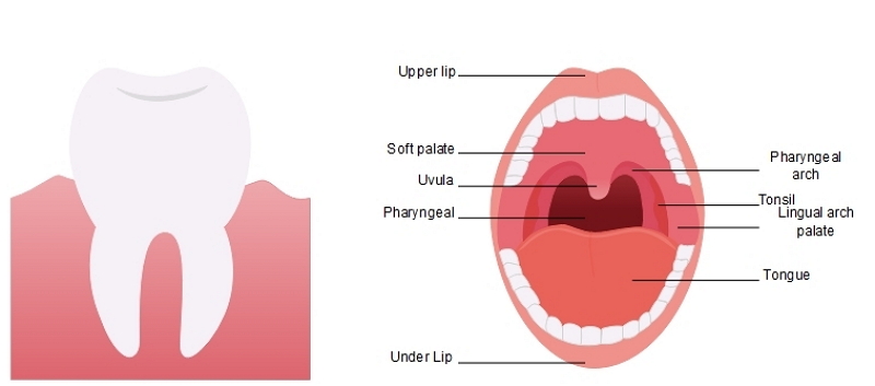 Source: EdrawMax Online
3. Teeth Structure and Functions
A tooth diagram is beneficial for learning about tooth anatomy. The teeth types present in the jaws have different shapes, but each tooth has a basic structure.
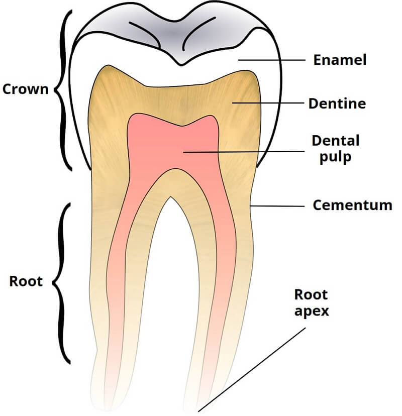
The apparent view of a tooth reveals only the crown portion. The part that is inside the gingiva or gum is called the root. An enamel layer protects the crown of the teeth. The root is protected from damage by the cementum. The layer separating the crown from the root is called the neck.
A detailed description of each part of a tooth include:
- Enamel: This is the layer covering the crown of a tooth. It functions as an insulator for a tooth. The tooth is protected from damage by the enamel. But the enamel can get damaged due to caries.
- Gingiva: This is the soft tissue surrounding the crown of the teeth. It helps in keeping the tooth in place. It also helps in protecting teeth from bacteria.
- Dentine: This layer is present beneath the enamel and cementum. It is the innervated tissue that forms the solid structure of a tooth.
- Pulp chamber: A space that is present beneath the dentine layer, which encapsulates the dental pulp. The dental pulp is the soft tissue that contains nerves and blood vessels.
- Cementum: It is the soft tissue that becomes visible due to a traumatic dental wound. The tooth is anchored with the alveolar bone and fibers with the help of the cementum.
- Root canal: It is a space that protects the pulp tissue of a tooth.
- Periodontal ligament: It is a connective tissue present between the tooth and the alveolar bone. A periodontal ligament helps in connecting the tooth to the jaw. The prime function of this ligament is to help the tooth resist the force of mastication.
- Accessory canal: It emerges as a branch of the primary root canal, offering access to the primary root canal.
- Apical foramen: It is a space in the apex of the tooth. Blood vessels and nerves enter the dental pulp through the apical foramen.
- Alveolar Bone: It is perforated and known as a cribriform plate, encircling the root of a tooth. Interalveolar nerves & vessels connect the periodontal ligament. Perforations help in this connection.
4. How to Draw a Tooth Diagram
Students can draw a tooth diagram to depict tooth anatomy. Practicing tooth diagrams helps in understanding the layers present in a tooth and also their functions. A lateral cross-section of a tooth is suitable for a diagram.
4.1 How to Draw Tooth Diagram from Sketch
Step 1: Students need to start from the crown and then draw the underlying layers one by one. The top layer is enamel and the second layer is dentine. The pulp chamber is inside the dentine, and the innermost part is the alveolar bone.
Step 2: After drawing these layers, the students need to create those parts that surround the tooth. It includes the gingiva and the periodontal ligament.
4.2 How to Draw Tooth Diagram Online
However, drawing a lateral cross-section of a tooth on paper is quite challenging. There is a second method of drawing a tooth diagram with the help of EdrawMax Online. Students can smoothly complete scientific diagrams and save those for further study with this tool. Follow the given steps to complete a tooth diagram with the EdrawMax Online tool:
Step 1: Open New on EdrawMax Online tool and click on Science Illustration or Science and Education.

Step 2: Choose Human Organ or Human Anatomy from the menu and customize the diagram. You will get many images related to the selected human organ.

Step 3: Once the students have selected their diagrams, they can smoothly work on them. They can modify the diagram according to their choice. It can help them to get a high-quality neuron labeled diagram apt for their study projects.
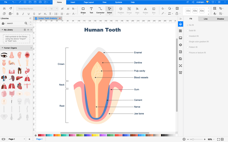
Step 4: Save the tooth diagram for further studies in tooth anatomy. You have the option to export the saved tooth diagram in any format, like the following image shows.

5. Tips for Healthy Teeth
It is essential to ensure good dental hygiene. A few simple steps are there to maintain healthy gums and teeth.
- Brush teeth twice a day
- Floss for trapped food particles
- Clean the tongue with a tongue cleaner
- Use mouthwash for killing bacteria
- Rinse mouth thoroughly after eating food
- Visit the dentist to assess dental health
6. Conclusion
Teeth are vital for ensuring proper digestion of food. Dental health is significant for leading a quality life. Students need to learn the importance of teeth health to prevent untimely teeth damage. They can view tooth diagrams at EdrawMax Online to enhance their knowledge.
In conclusion, EdrawMax Online is a quick-start diagramming tool, which is easier to make artery and vein diagram and any 280 types of diagrams. Also, it contains substantial built-in templates that you can use for free, or share your science diagrams with others in our template community.


