A Guide to Understand Mucor with Diagram
Mucor is mainly available on soil, rotten food, but they can be the reason for infection in human bodies and other amphibians. It is essential to know the anatomy and life cycle of Mucor to understand its nature and the diseases. For a thorough understanding, the students can use a Mucor diagram. However, it is challenging to create a Mucor diagram by hand, and for that, the students must use the EdrawMax Online tool.
1. What is the Mucor
Mucor is a fungus that is universally present. They are fast-growing and can affect the lungs, eyes, skin, and mucous membrane. About a total of 80 species, there are 17 of them available in India. They are saprophytic colonized by nature. The Mucor uses simple carbohydrate molecules and then utilizes complex carbohydrates.

1.1 The Mucor Species
Mucor is a fungus that belongs to the Zygomycota family. They are fast-growing and primarily available in the soil, stale food, and rotting vegetables. They are also responsible for some infectious reactions in man, cattle, frogs, and other amphibians. There are about 50 species of Mucor that are found all over the world. Some common Mucor species are amphibious, circinelloides, indicus, racemosus, hiemalis, ramosissimus, etcetera.
1.2 The Structure of Mucor
Mycelium: Network of hyphae forms A mycelium. They are branched and create a network-like structure made of hyphae.
Hyphae: Mucor is formed with a filamentous, thin structure known as hyphae. These thin, thread-like structures help in creating a network. There are mainly three types of hyphae:
-
Subterranean hyphae:
- They are highly branched.
- They are located horizontally to the substratum.
- They help to absorb water and nutrients.
- Prostrate hyphae: They are located horizontally under or between substratum and help to absorb water and nutrients.
- Aerial hyphae: They are present vertically on the prostrate hyphae.
- Sporangiophore: Sporangiophores are thin and long in structure. These structures work to make and store spores in the Mucor.
- Columella: Columella has varied shapes and sizes. The sporangiophores swell up to form a column-shaped structure, known as the columella. These sterile dome-shaped structures participate in the nutritional exchange between the protoplasm of the developing spores and sporangial heads.
- Sporangium: It is a thick covering that contains numerous spores. They can be spherical or globular in shape, and columella, the column-like structure, supports them. They participate in asexual reproduction.
- Spores: The sporangium comes in various shapes, and they are flattened. This thick covering of the sporangium has spores. They are the reproductive structures present in the Mucor.
- Nucleus: Mucor has small multinucleated nuclei.
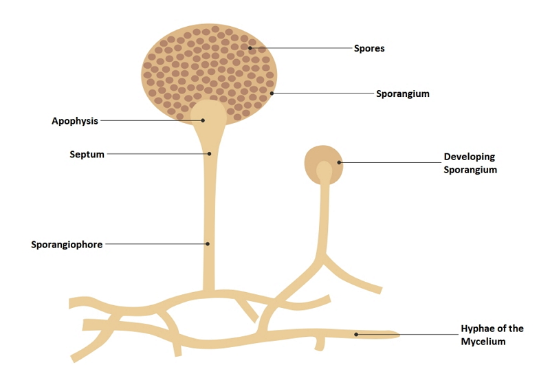 Source:EdrawMax Online
2. How to Draw the Mucor Diagram?
A Mucor diagram can help a student to have an apt idea about the same. Mucor shows three types of reproductive features, which include spores. Therefore, knowing about the anatomy also helps in understanding the life cycle. The students can also have an idea about the infections caused by it. The student may use a Mucor diagram to study the different parts of Mucor.
2.1 How to Create Mucor Diagram from Sketch
The students can create the diagram by hand, but the process is lengthy and complicated. They need to follow these steps to draw the same:
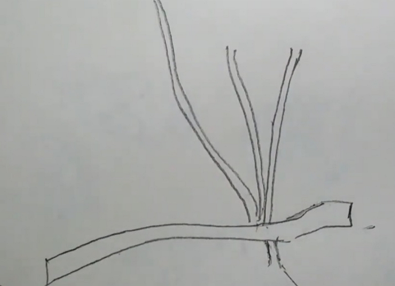
Step 1: First, the student needs to draw a curved line and make a double line. After that, they should join the ends. There are uneven structures present within these curved shapes, which are called the Hyphae.

Step 2: Then they need to draw some column-like structure with several branches coming out of the curved structure. The column-like structures are known as Sporangiophores.
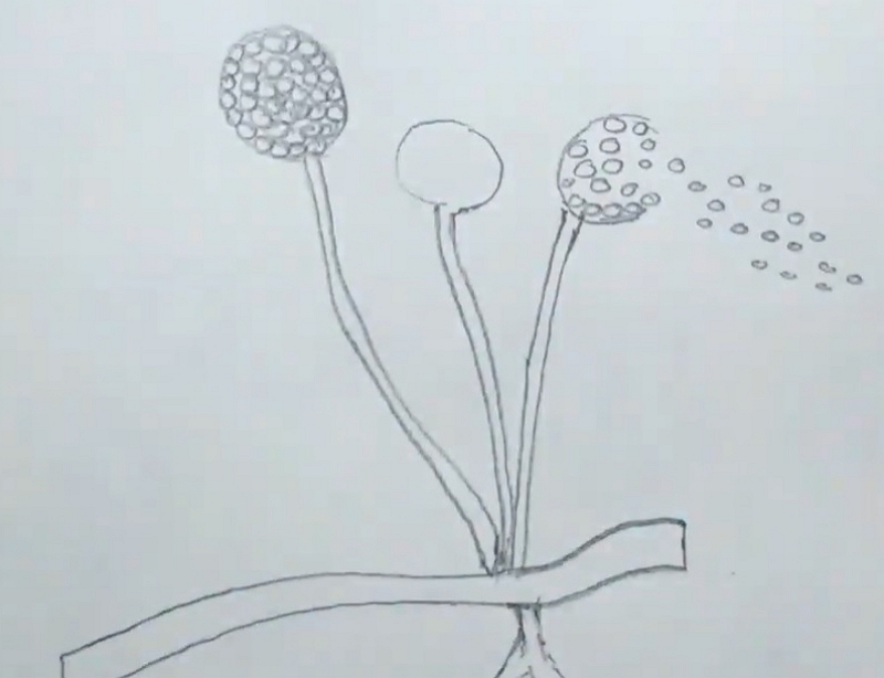
Step 3: Draw a swollen, circular structure at the top of the column-like figures. These are known as sporangia. Small circular shapes are covering the circular top of the Mucor and are the spores of the same.
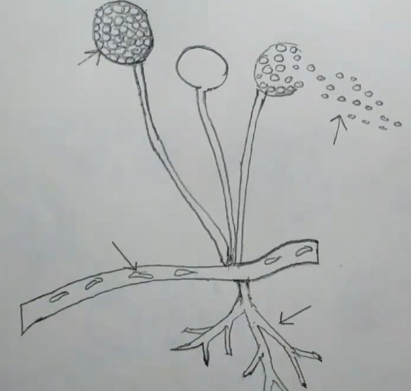
Step 4: The bottom part of the column forms a root-like structure that has several branches. These are called Rhizoids.
2.2 How to Create Mucor Diagram Online
Though the students can draw a Mucor diagram by hand, the process is complicated and may take time. The students may fail to get a satisfactory result. To avoid such hassle, the students must use the EdrawMax Online tool. The tool is user-friendly, and hence any inexperienced user can comfortably work on this tool. Here are a few simple steps which they need to follow to create an easy Mucor diagram with the help of the EdrawMax Online tool:
Step 1: EdrawMax Online tool has an easy-to-use interface which makes it comfortable for the users to work on this tool. To start with the process, the students need to open the EdrawMax Online tool. It is a powerful diagramming tool and trusted by many individuals as their diagramming partner. The students have to open New and then look for the Science and Education Option.

Step 2: The students can use this tool for creating different types of science and education-related diagrams. EdrawMax Online tool provides the students with high-quality images that are perfect for their lessons and projects. They can be time-saving for the students as well. There is the Biology option present under the Science and Education tab.

Step 3: In the Biology option, there are multiple biology-related diagrams present for the students. The students can look for the Mucor diagram. These diagrams are a perfect fit for projects and dissertation papers. The students can easily edit those pictures as per their requirements and use them for their lessons.
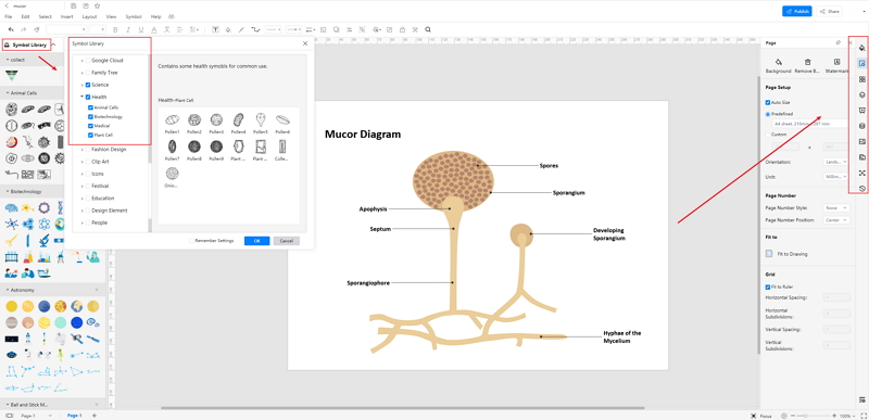
Step 4: Once the student is satisfied with their diagram, they can save it. EdrawMax Online tool allows the students to Save it in multiple file formats. They can also export it for future use. Finally, the diagram is ready for them to use in projects or for their studies.

3. Life Cycle of Mucor
Mucor is the type of fungi that is available worldwide. It has three different types of reproduction, which help it to fulfill its life cycle. They are:
Vegetative reproduction: The vegetation reproduction of the Mucor occurs through fragmentation. The fragments of a vegetative cell form new bodies in favorable conditions.
Asexual reproduction: Non-motile spores like sporangiospores, chlamydospores, and Oidiospores participate in asexual reproduction. For sporangiospores, an unbranched sporangiophore arises. After maturation, the aerial hyphae get swollen and forms a large sporangium. While developing, the sporangium gets separated into sporoplasm and columella plasma. The cell lysis occurs, and then they reproduce a new Mucor. The Oidiospores remain dormant for a particular time before they get favorable conditions. They create germination tubes to create a new vegetative body.
Sexual reproduction: The sexual reproduction in Mucor occurs through Gametangial conjugation. Thallus of two opposite strains come in contact and then form a Progametangium. The gametes form zygote. The zygospores stay dormant until favorable conditions. After that, promycelium develops to create a new vegetative body.
4. Conclusion
To know about Mucor, the students must learn about their anatomy, species, and reproductive methods. The students can use Mucor diagrams to learn about them. However, creating them by hand is difficult and time-consuming. The students must use the EdrawMax Online tool, which is user-friendly, and the students can create a high-quality Mucor diagram without much trouble.
In conclusion, EdrawMax Online is a quick-start diagramming tool, which is easier to make artery and vein diagram and any 280 types of diagrams. Also, it contains substantial built-in templates that you can use for free, or share your science diagrams with others in our template community.


